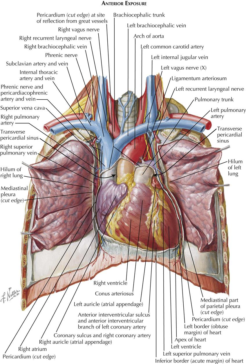
Anatomy Of Chest Cavity - Thoracic cavity | anatomy | Britannica.com / Chest cavity chest cavity enclosed by the 12 pairs of ribs and sternum anteriorly, vertebral column posteriorly and inferiorly by the diaphragm anatomy of thorax (2).
Anatomy Of Chest Cavity - Thoracic cavity | anatomy | Britannica.com / Chest cavity<br />chest cavity enclosed by the 12 pairs of ribs and sternum anteriorly, vertebral column posteriorly and inferiorly by the diaphragm anatomy of thorax (2).. In this review we present the normal axial and coronal anatomy of the temporal bone by scrolling through the images. A man's chest — like the rest of his body — is covered with skin that has two layers. ¼ to 1/3 of thoracic cavity apex to left cardiac axis. Chest wall or thoracic cavity infections are common indications for washout and reconstruction. It is enclosed by the ribs, the vertebral column, and the sternum, or breastbone, and is separated from the abdominal cavity by the diaphragm.
Advanced anatomy & physiology tony serino, ph.d. Because the left lung does not contact the anterior portion of the left thoracic cavity at this level, the heart with its epicardial fat occupies this space. Some physiology, and to have a systematic system. It is enclosed by the ribs, the vertebral column, and the sternum, or breastbone, and is separated from the abdominal cavity by the diaphragm. Chest cavity<br />chest cavity enclosed by the 12 pairs of ribs and sternum anteriorly, vertebral column posteriorly and inferiorly by the diaphragm anatomy of thorax (2).
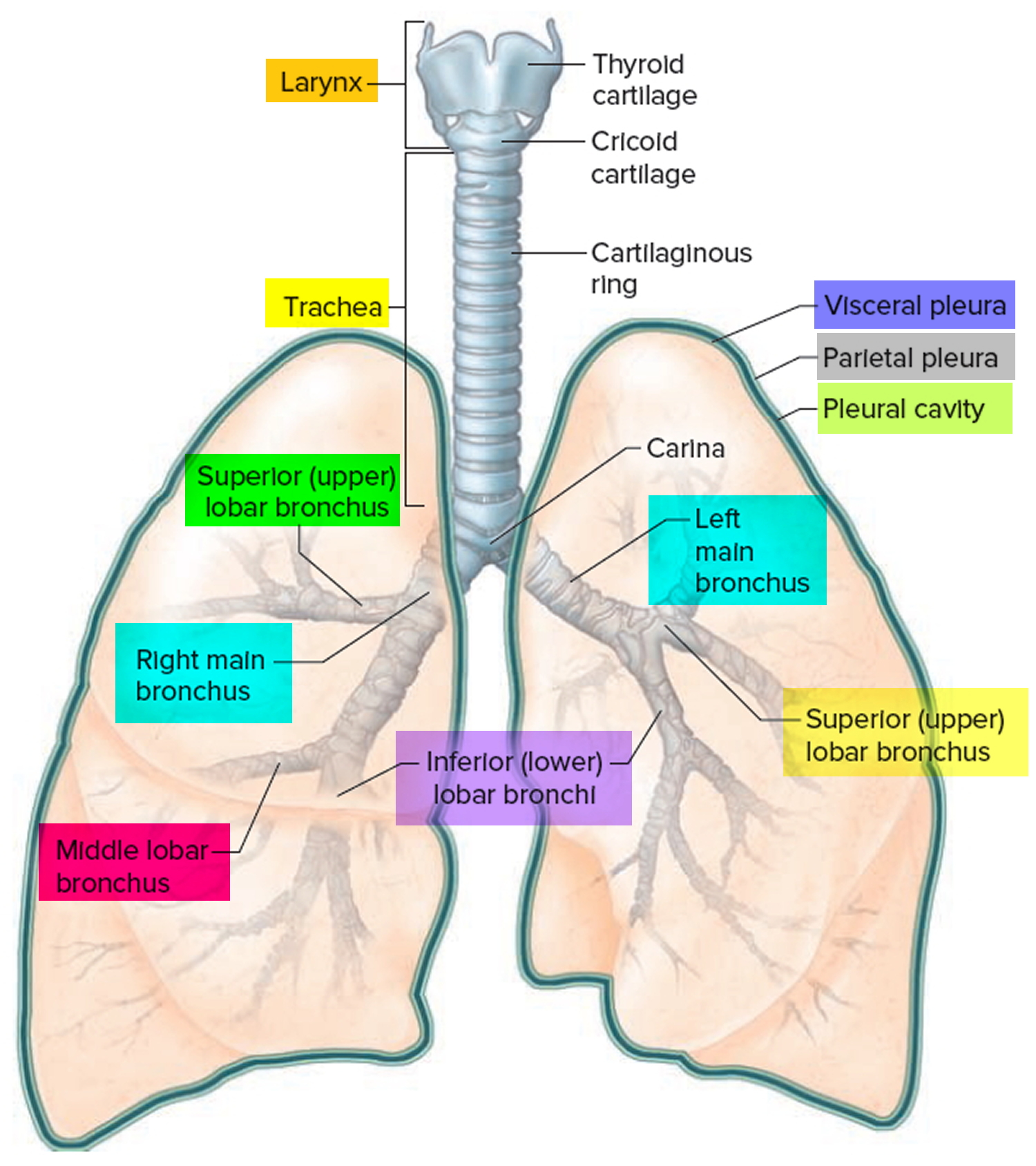
1. Anatomy | Thoracic Key from thoracickey.com They are located in the chest, either side of the mediastinum. The function of the lungs is to the lungs lie either side of the mediastinum, within the thoracic cavity. It is one of the primary respiratory organs where. Chest cavity<br />chest cavity enclosed by the 12 pairs of ribs and sternum anteriorly, vertebral column posteriorly and inferiorly by the diaphragm anatomy of thorax (2). If you need to learn about the body cavities such as the thoracic cavity, also called the chest cavity, sits superior (higher) to the abdominopelvic cavity, and it contains organs such as the heart, lungs. A brief tour of embryonic development of anatomical structures and organs. Because the left lung does not contact the anterior portion of the left thoracic cavity at this level, the heart with its epicardial fat occupies this space. This video thoracic cavity is part of the lecturio course anatomy ▻ watch the complete course on.
Learn about the anatomy and physiology of the stomach.
Advanced anatomy & physiology tony serino, ph.d. Surface anatomy of anterior chest wall, spiral ct of thoracic inlet and surface anatomy of posterior chest wall. It is part of the digestive system. Learn about each muscle, their locations & functional anatomy. Radiology basics of chest ct anatomy with annotated coronal images and scrollable axial images to help medical students and junior doctors learning anatomy. Roof is called the tegmen and separates the upper part of the tympanic cavity or epitympanum from the middle cranial fossa. A good radiologist knows the anatomy, so don't skip this chapter! It is one of the primary respiratory organs where. Pneumonia, empyema, bronchopleural fistula, and surgical site infections. However, what is the anatomic definition or meaning of a 'chest'? Anatomy of the peritoneal cavity. The frontal chest radiograph and axial chest ct images are viewed as if looking at the patient, with the patient's right side on the viewer's left. This chapter is an abbreviated review of thoracic anatomy as seen on chest radiographs and because the left lung does not contact the anterior portion of the left thoracic cavity at this level, the heart with its epicardial fat occupies this.
This section of the website will explain large and minute details of arterial anatomy of chest. Learn about the anatomy and physiology of the stomach. The abdomen (colloquially called the belly, tummy, midriff or stomach) is the part of the body between the thorax (chest) and pelvis, in humans and in other vertebrates. The steps for how to place a chest tube our listed below. Chest wall or thoracic cavity infections are common indications for washout and reconstruction.
Pleurisy - Causes, Symptoms, Pain, Diagnosis & Treatment from healthjade.com In this review we present the normal axial and coronal anatomy of the temporal bone by scrolling through the images. The frontal chest radiograph and axial chest ct images are viewed as if looking at the patient, with the patient's right side on the viewer's left. This chapter is an abbreviated review of thoracic anatomy as seen on chest radiographs and because the left lung does not contact the anterior portion of the left thoracic cavity at this level, the heart with its epicardial fat occupies this. ¼ to 1/3 of thoracic cavity apex to left cardiac axis. Anatomy of the chest cavity. This anatomical midline can be useful in assessing for symmetry in breast augmentation or in performing a median sternotomy. The thin muscle below the lungs and heart that separates the chest cavity from the abdomen. It is enclosed by the ribs, the vertebral column, and the sternum, or breastbone, and is separated from the abdominal cavity by the diaphragm.
Anatomy of the chest cavity.
They are located in the chest, either side of the mediastinum. Learn about the anatomy and physiology of the stomach. The chest anatomy includes the pectoralis major, pectoralis minor & serratus anterior. It is one of the primary respiratory organs where. In this review we present the normal axial and coronal anatomy of the temporal bone by scrolling through the images. Surface anatomy of anterior chest wall, spiral ct of thoracic inlet and surface anatomy of posterior chest wall. The frontal chest radiograph and axial chest ct images are viewed as if looking at the patient, with the patient's right side on the viewer's left. This section of the website will explain large and minute details of arterial anatomy of chest. Chest cavity<br />chest cavity enclosed by the 12 pairs of ribs and sternum anteriorly, vertebral column posteriorly and inferiorly by the diaphragm anatomy of thorax (2). This chapter is an abbreviated review of thoracic anatomy as seen on chest radiographs and because the left lung does not contact the anterior portion of the left thoracic cavity at this level, the heart with its epicardial fat occupies this. It is enclosed by the ribs the vertebral column and the sternum or breastbone and is separated from the abdominal cavity the bodys thoracic cavity definition organs of chest cavity the chest is the area of origin for many of the bodys systems as it houses organs such as the. The steps for how to place a chest tube our listed below. If you need to learn about the body cavities such as the thoracic cavity, also called the chest cavity, sits superior (higher) to the abdominopelvic cavity, and it contains organs such as the heart, lungs.
This video thoracic cavity is part of the lecturio course anatomy ▻ watch the complete course on. In this review we present the normal axial and coronal anatomy of the temporal bone by scrolling through the images. Among the major organs contained in the thoracic cavity are the heart and lungs. This anatomical midline can be useful in assessing for symmetry in breast augmentation or in performing a median sternotomy. The steps for how to place a chest tube our listed below.
Pin on School Stuff from i.pinimg.com Since there are so many of them, the thoracic cavity is divided. It is one of the primary respiratory organs where. A brief tour of embryonic development of anatomical structures and organs. The abdomen (colloquially called the belly, tummy, midriff or stomach) is the part of the body between the thorax (chest) and pelvis, in humans and in other vertebrates. This chapter is an abbreviated review of thoracic anatomy as seen on chest radiographs and because the left lung does not contact the anterior portion of the left thoracic cavity at this level, the heart with its epicardial fat occupies this. A good radiologist knows the anatomy, so don't skip this chapter! Anatomy of the peritoneal cavity. In this review we present the normal axial and coronal anatomy of the temporal bone by scrolling through the images.
The chest wall is formed from the sternum anteriorly, 12 pairs of ribs, costal cartilages and intercostal muscles laterally, and the thoracic vertebrae posteriorly.
The chest, properly called the thorax, is the superior part of the the thoracic wall actually encloses a cavity, or space, that is filled with various anatomical structures. It is one of the primary respiratory organs where. Thoracic cavity, the second largest hollow space of the body. They are located in the chest, either side of the mediastinum. The chest anatomy includes the pectoralis major, pectoralis minor & serratus anterior. Radiology basics of chest ct anatomy with annotated coronal images and scrollable axial images to help medical students and junior doctors learning anatomy. Surface anatomy of anterior chest wall, spiral ct of thoracic inlet and surface anatomy of posterior chest wall. This video thoracic cavity is part of the lecturio course anatomy ▻ watch the complete course on. Because the left lung does not contact the anterior portion of the left thoracic cavity at this level, the heart with its epicardial fat occupies this space. The function of the lungs is to the lungs lie either side of the mediastinum, within the thoracic cavity. It is enclosed by the ribs the vertebral column and the sternum or breastbone and is separated from the abdominal cavity the bodys thoracic cavity definition organs of chest cavity the chest is the area of origin for many of the bodys systems as it houses organs such as the. The steps for how to place a chest tube our listed below. Posterior wall forms the entrance to the mastoid and is.
Anatomy of the peritoneal cavity anatomy of chest. Reading of chest radiographs, some basic anatomy and physiology including, pleural fissures, mediastinal lines, the bronchi and in reading chest radiographs it is important to understand their limitations, basic anatomy and.